Hammer Toe Repair
 Overview
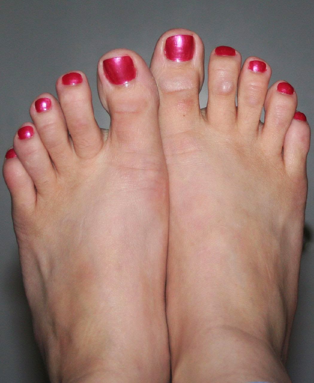
hammertoes deformities can be painful and unsightly. These toe deformities can be the result of a muscle/tendon imbalance or often the end stage result of some systemic disease such as diabetes or arthritis, especially Rheumatoid arthritis. Hammertoe deformities are progressive and can be prevented.
Causes
People who have a high-arched feet have an increased chance of hammer toes occurring. Also, patients with bunion deformities notice the second toe elevating and becoming hammered to make room for the big toe that is moving toward it. Some patients damage the ligament that holds the toe in place at the bottom of the joint that connects the toe and foot. When this ligament (plantar plate) is disrupted or torn, the toe floats upward at this joint. Hammer toes also occur in women wearing ill-fitting shoes or high heels, and children wearing shoes they have outgrown.
 Symptoms
The most obvious symptoms of this injury will be the the middle toe joint is permanently bent at an angle. In the beginning movement may still be possible but as time passes and the injury worsens the toe will be locked in place and possible require hammer toe correction surgery to fix. Another key indicator of hammer toe is that a lump or corn will form on top of the toe. The toe joint will be painful and walking can cause severe discomfort. Occasionally a callus may form on the sole of the injured foot. If you see any of these symptoms together or have been enduring pain for some time, seeing a podiatrist should be your next step.
Diagnosis
Your doctor is very likely to be able to diagnose your hammertoe simply by examining your foot. Even before that, he or she will probably ask about your family and personal medical history and evaluate your gait as you walk and the types of shoes you wear. You'll be asked about your symptoms, when they started and when they occur. You may also be asked to flex your toe so that your doctor can get an idea of your range of motion. He or she may order x-rays in order to better define your deformity.
Non Surgical Treatment
If the problem is caught in the early stages you can avoid hammer toe surgery. One of the easiest methods of treatment is to manipulate the toe out of a bent position then splint and buddy wrap it alongside it?s larger neighbour. This Hammer toe method of hammer toe taping will help the problem to fix itself. Make sure the toe isn?t resuming its bent shape during the recovery. To alleviate some of the painful symptoms of hammer toe avoid wearing high heels or shoes that cramp or stifle your feet. Choosing a pair of minimalist shoes can be an excellent choice for both foot and postural health. Wearing shoes that give the toes plenty of space and are comfortable lined is also a smart choice. Hammer toe recovery starts be treating the toe respectfully. Soft insoles or protection for the corn can also provide additional assistance.
Surgical Treatment
If conservative measures fail to provide relief, or if your hammertoe is in advanced stages with rigidity and a significant amount of pain, surgery may be required. Some patients also require surgery if they have open sores or wounds related to their hammertoe. For patients who also suffer from bunions, a combined procedure may be appropriate, addressing both conditions within the same surgery. Recovery time will vary from patient to patient, depending on the extent of the surgical repair and other conditions that may also be present.
What Are The Treatments For Bunions?
Overview
 The term hallux valgus actually describes what happens to the big toe. Hallux is the medical term for big toe, and valgus is an anatomic term that means the deformity goes in a direction away from the midline of the body. So in hallux valgus the big toe begins to point towards the outside of the foot. As this condition worsens, other changes occur in the foot that increase the problem. One of those changes is that the bone just above the big toe, the first metatarsal, usually develops too much of an angle in the other direction. This condition is called metatarsus primus varus. Metatarsus primus means first metatarsal, and varus is the medical term that means the deformity goes in a direction towards the midline of the body. This creates a situation where the first metatarsal and the big toe now form an angle with the point sticking out at the inside edge of the ball of the foot. The bunion that develops is actually a response to the pressure from the shoe on the point of this angle. At first the bump is made up of irritated, swollen tissue that is constantly caught between the shoe and the bone beneath the skin. As time goes on, the constant pressure may cause the bone to thicken as well, creating an even larger lump to rub against the shoe.
Causes
Perhaps the most frequent cause of bunion development is the wearing of shoes with tight, pointed toes, or with high heels that shift all of your body's weight onto your toes and also jam your toes into your shoes' toe boxes. It's estimated that more than 50 percent of women have bunions caused by high-heel shoes, and that nine out of 10 people who develop bunions are women. Bunions can also develop on your little toes, in which case they are called bunionettes or tailor's bunions.
Symptoms
No matter what stage your bunion is in, you can be in pain. Though bunions take years to develop, you can experience pain at any stage. Some people don?t have bunion pain at all. Pain from a bunion can be severe enough to keep you from walking comfortably in normal shoes. The skin and deeper tissue around the bunion also may become swollen or inflamed.
Diagnosis
A thorough medical history and physical exam by a physician is necessary for the proper diagnosis of bunions and other foot conditions. X-rays can help confirm the diagnosis by showing the bone displacement, joint swelling, and, in some cases, the overgrowth of bone that characterizes bunions. Doctors also will consider the possibility that the joint pain is caused by or complicated by Arthritis, which causes destruction of the cartilage of the joint. Gout, which causes the accumulation of uric acid crystals in the joint. Tiny fractures of a bone in the foot or stress fractures. Infection. Your doctor may order additional tests to rule out these possibilities.
Non Surgical Treatment
Podiatrists will treat bunions conservatively, using paddings and orthotics, which are devices that are made to protect the joint or deviate pressure away from it. Sometimes bunions will develop overlying callus or corns. These can be removed by a podiatrist, but if the area is irritated again by wearing ill-fitting footwear, the corn will grow back. Most people with this condition have flat feet, so arch supports are often recommended. 
Surgical Treatment
Larger bunions are commonly treated with the Lapidus Bunionectomy, which involves realigned the displaced bone at its bottom, through a bone mending/fusion procedure. It is with this technique where the the walking advances have been made int he past decade. The Lapidus Bunionectomy has become a popular surgical method since surgeons have become more comfortable with mobilizing their patients post-operatively. Though not all surgeons who perform this procedure have adopted this postoperative protocol.
Pain In The Arch Of My Foot And Heel
Overview
Pes planus is the scientific term that describes low arch or "flat" feet. As podiatric physicians, we are very interested not only in structure but also in function of the feet. While very few people have 20-20 vision, it is also true that very few people have perfect arch structure. High and low arch feet are just the two ends of the spectrum of foot structure. The more deviation from what is considered perfect, the worse the function becomes. We describe deviation from "the ideal" as imbalance in structure. It is the imbalance in structure that leads to abnormal function. Abnormal function causes pain and/or deformity. Deformity can manifest itself in a variety of foot problems such as bunions or hammertoes. Likewise, pain can manifest itself in a variety of ways such as heel pain ( plantar fasciitis or heel spur), corns or calluses, metatarsalgia or pain in the ball of the foot ( neuromas, stress fractures, or tendonitis), or even in pediatric problems. Invariably, we can treat most foot imbalance or biomechanical insufficiencies with orthotic therapy. 
Causes
The cause of this condition is too much pressure exerted on the arches, and although common in athletes, the condition can happen because you went hiking or climbing, you were lifting heavy objects, or you simply walked too far too vigorously. Pregnancy places extra strain on the arches because of both the additional body weight and the effect of hormones on muscles and ligaments.
Symptoms
Flat feet can exhibit a variety of symptoms, from mild to severe. The extent of the flat foto does not always correlate with the extent of symptoms. Patients may complain of arch pain and heel pain. Commonly there is pain on the outside of the foot, where the foot meets the ankle as the collapse foot abuts against the ankle. Muscle cramps within the foot, and onto the leg (shin splints) may occur. In general, patients have pain with activity, such as walking or running. The pain may be deep and focal to a generalized widespread achy feeling. Irritation from shoe gear can cause redness and swelling. Common reasons patients seek treatment are pain, interference with walking or activities, difficulty fitting shoes, swelling, and notice a change in appearance of the foot and/or unsightly appearance.
Diagnosis
Your doctor may order imaging tests to help make sure your heel pain is caused by plantar fasciitis and not another problem. X-rays provide clear images of bones. They are useful in ruling out other causes of heel pain, such as fractures or arthritis. Heel spurs can be seen on an x-ray. Other imaging tests, such as magnetic resonance imaging (MRI) and ultrasound, are not routinely used to diagnose plantar fasciitis. They are rarely ordered. An MRI scan may be used if the heel pain is not relieved by initial treatment methods.
Non Surgical Treatment
Rest from any aggravating activities and apply cold therapy whilst in the acute phase when the foot or ankle is painful and inflamed. Your Doctor may prescribe anti-inflammatory medication to help reduce pain and inflammation. Once the initial pain and inflammation has gone then a full rehabilitation program which includes stretching and strengthening exercises for the foot can begin. Consult a foot specialist, podiatrist or physiotherapist who can assess this and biomechanical problems of the foot. Nerve conduction studies may be done to confirm the diagnosis and indicate the location of the entrapment. An X-ray or MRI may also be useful to determine the presence of any other structures such as cysts, arthritis or a tarsal coalition. 
Surgical Treatment
If you have pain that has not been responsive to other treatments, there is a new non-surgical treatment that was recently approved by the FDA. ESWT (extracorporeal shockwave therapy) uses strong electrohydraulic acoustic (sound) energy that triggers the body?s natural repair mechanism. This treatment method is safe, effective and requires a very short recovery period compared to older surgical techniques.
Prevention
Strap the arches into the anatomically correct positions with athletic tape and leave them like this for some time. If the fallen arches are an issue with the muscular structure, this may give the muscles an opportunity to strengthen. This is definitely not a fallen arches cure all the time but it can help prevent it more times than not. Ask a doctor or physical therapists to show you how to do this taping. Find shoes that fit. This may require that you get your foot measured and molded to ensure that the shoe will fit. Shoes that are too big, too tight or too short, may not directly cause the fallen arches, but they can assist with the damage to the area. These shoes should have thick cushioning inside and have plenty of room for your toes. Walk without shoes as much as possible. Shoes directly assist with weakening and distorting the arches of the feet so going without shoes can actually help strengthen your arches and prevent fallen arches. Walking on hard and bumpy surfaces barefooted makes the muscles in your feet strengthen in order to prevent injury. It is a coping mechanism by your body. Insert heel cups or insoles into the shoes that you wear the most. Many people wear uncomfortable shoes to work and these are the same shoes that cause their arches the most problems. Inserting the heel cups and insoles into these shoes can prevent fallen arches from occurring. Many people place these inserts into all their shoes to ensure support. Ask a medical professional, either your doctor or a physical therapist, about daily foot exercises that may keep the arches stronger than normal. Many times, you can find exercises and stretches on the Internet on various websites. Curling your toes tightly and rotating your feet will help strengthen your longitudinal arches. Relax your feet and shake them for a minute or so before you do any arch exercises. This will loosen the muscles in your feet that stay tight due to normal daily activities. Wear rigid soled sandals whenever possible to provide a strong support for your arches. Wooden soled sandals are the best ones if available. Walk or jog on concrete as much as you can. This will create a sturdy support for your arches. Running or walking in sandy areas or even on a treadmill, does not give rigid support. Instead, these surfaces absorb the step, offering no support for arches.
Stretching Exercises
Achilles stretch. Stand with the ball of one foot on a stair. Reach for the step below with your heel until you feel a stretch in the arch of your foot. Hold this position for 15 to 30 seconds and then relax. Repeat 3 times. Balance and reach exercises. Stand next to a chair with your injured leg farther from the chair. The chair will provide support if you need it. Stand on the foot of your injured leg and bend your knee slightly. Try to raise the arch of this foot while keeping your big toe on the floor. Keep your foot in this position. With the hand that is farther away from the chair, reach forward in front of you by bending at the waist. Avoid bending your knee any more as you do this. Repeat this 15 times. To make the exercise more challenging, reach farther in front of you. Do 2 sets of 15. While keeping your arch raised, reach the hand that is farther away from the chair across your body toward the chair. The farther you reach, the more challenging the exercise. Do 2 sets of 15. Towel pickup. With your heel on the ground, pick up a towel with your toes. Release. Repeat 10 to 20 times. When this gets easy, add more resistance by placing a book or small weight on the towel. Resisted ankle plantar flexion. Sit with your injured leg stretched out in front of you. Loop the tubing around the ball of your foot. Hold the ends of the tubing with both hands. Gently press the ball of your foot down and point your toes, stretching the tubing. Return to the starting position. Do 2 sets of 15. Resisted ankle dorsiflexion. Tie a knot in one end of the elastic tubing and shut the knot in a door. Tie a loop in the other end of the tubing and put the foot on your injured side through the loop so that the tubing goes around the top of the foot. Sit facing the door with your injured leg straight out in front of you. Move away from the door until there is tension in the tubing. Keeping your leg straight, pull the top of your foot toward your body, stretching the tubing. Slowly return to the starting position. Do 2 sets of 15. Heel raise. Stand behind a chair or counter with both feet flat on the floor. Using the chair or counter as a support, rise up onto your toes and hold for 5 seconds. Then slowly lower yourself down without holding onto the support. (It's OK to keep holding onto the support if you need to.) When this exercise becomes less painful, try doing this exercise while you are standing on the injured leg only. Repeat 15 times. Do 2 sets of 15. Rest 30 seconds between sets.
Pain In The Arches All You Need To Learn
Overview
Pes planus is the scientific term that describes low arch or "flat" feet. As podiatric physicians, we are very interested not only in structure but also in function of the feet. While very few people have 20-20 vision, it is also true that very few people have perfect arch structure. High and low arch feet are just the two ends of the spectrum of foot structure. The more deviation from what is considered perfect, the worse the function becomes. We describe deviation from "the ideal" as imbalance in structure. It is the imbalance in structure that leads to abnormal function. Abnormal function causes pain and/or deformity. Deformity can manifest itself in a variety of foot problems such as bunions or hammertoes. Likewise, pain can manifest itself in a variety of ways such as heel pain ( plantar fasciitis or heel spur), corns or calluses, metatarsalgia or pain in the ball of the foot ( neuromas, stress fractures, or tendonitis), or even in pediatric problems. Invariably, we can treat most foot imbalance or biomechanical insufficiencies with orthotic therapy. 
Causes
Tarsal Tunnel Syndrome develops when there is compression on the tibial nerve as it passes through the tarsal tunnel on the inner side of the ankle bone (medial malleolus). It can cause pain on bottom of foot as well as pins and needles. Numbness in the heel can often extend down to the big toe and adjacent three toes. In addition, it may also produce hot and cold sensations along the bottom of the foot. Tarsal Tunnel Syndrome is caused by anything which occupies space in the tarsal tunnel including cysts, ganglions, bone spurs, swelling from ankle injuries or tumours. Treatment aims to reduce the foot arch pain and usually consists of rest, strengthening and stretching exercises, compression bandages and steroid injections. If the pain in bottom of foot persists, surgery may be required.
Symptoms
Most flat feet do not cause pain or other problems. Children may have foot pain, ankle pain, or lower leg pain. They should be evaluated by a health care provider if this occurs. Symptoms in adults may include tired or achy feet after long periods of standing or playing sports.
Diagnosis
Magnetic Resonance Imaging (MRI) can show tendon injury and inflammation but cannot be relied on with 100% accuracy and confidence. The technique and skill of the radiologist in properly positioning the foot with the MRI beam are critical in demonstrating the sometimes obscure findings of tendon injury around the ankle. Magnetic Resonance Imaging (MRI) is expensive and is not necessary in most cases to diagnose posterior tibial tendon injury. Ultrasound has also been used in some cases to diagnose tendon injury, but this test again is usually not required to make the initial diagnosis.
Non Surgical Treatment
If the strain is severe enough, it can not only stretch but tear the plantar fascia. No matter what the cause of your problem, however, the end result is the same, foot pronation, a temporary case of "flat feet" and pain. The best treatment? Apply ice packs, followed by heat (to reduce inflammation), to the area for 20 minutes once a day. Rest is also essential. You will have to avoid any activity, in some cases, even standing or walking, that would increase the tear, until the tissue heals on its own (this can sometimes take up to six weeks). With strains and less severe tears, you may be able to walk on the foot with arch-support shoe inserts. You'll need to see your doctor for more permanent arch support. A doctor can also provide immediate relief from the pain of plantar fasciitis by giving you a local cortisone injection or prescribing anti-inflammatory medication. 
Surgical Treatment
A procedure that involves placing a metallic implant (most commonly) at the junction where the foot meets the ankle. This device causes the physical blockade that prevent the collapse. It is a procedure that is only indicated for mobile feet, and should not be used with rigid flat feet. Dr. Blitz finds this procedure better for younger patients with flexible flat feet where the bone alignment is still developing so that the foot can adapt to function in a better aligned position.
Prevention
There are several things you can do to prevent pain on the bottom of the foot. Here are some tips to help you avoid this condition. Do simple stretches each day (See Plantar Fasciitis Exercises for a list of all exercises). Wear good shoes that fit properly and are appropriate for the activity you are participating in. Lose excess weight if possible. Build your stamina slowly, especially with new exercises. Rest and elevate your feet, whenever possible, keeping them at least twelve inches above your heart. Always follow your doctor?s instructions for treatment. Each day do a different activity. For example: one day ride your bike, and swim the next day.
Stretching Exercises
Below are two simple plantar fasciitis stretching exercises to help improve the flexibility of the muscles and tendons around the foot and ankle. Plantar fasciitis stretch taken from The Stretching Handbook. Kneel on one foot and place your body weight over your knee. Keep your heel on the ground and lean forward. In the photo to the left, the athlete is stretching the arch of her left foot. Kneel on one foot with your hands on the ground. Place your body weight over your knee and slowly move your knee forward. Keep your toes on the ground and arch your foot. In the photo to the right, the athlete is stretching the arch of his right foot.